Types of Tympanoplasty - Wullstein Classification
💎 Buy my Premium ENT Notes
Instant access to 200+ high-yield ENT notes. Your purchase includes all future updates.
🇮🇳 For Indian Students
· To buy all my notes, click here💡 This post is a free outline of my YouTube video. Get my full handwritten notes using the links above.
🦻 Types of Tympanoplasty – Wullstein Classification
Tympanoplasty is the surgical reconstruction of the hearing mechanism of the middle ear, typically due to chronic otitis media.
The most widely accepted tympanoplasty classification was proposed by Wullstein. It divides the procedure into five types based on:
- The site of defect
- The status of the ossicular chain
- Where the graft is placed
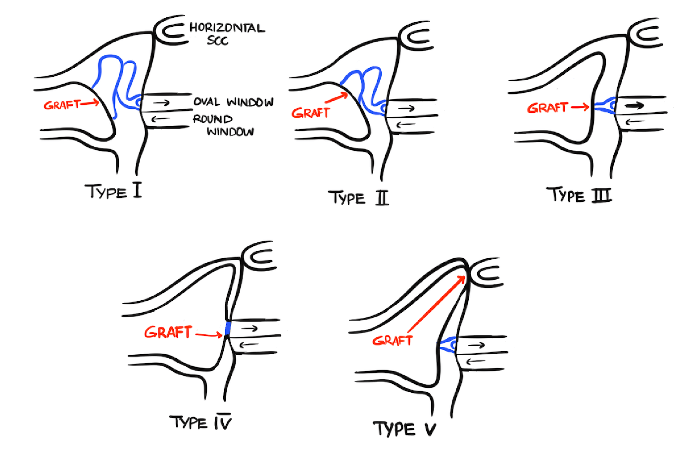
The diagram above illustrates the five types in Wullstein classification of tympanoplasty.
📘 Type 1 Tympanoplasty
- 🦴 Ossicles: Intact (Malleus, Incus, Stapes)
- 🕳️ Defect: Only tympanic membrane perforation
- 🩹 Graft Placement: On the remnant tympanic membrane or on the handle of malleus
📘 Type 2 Tympanoplasty
- 🦴 Ossicles: Malleus is eroded, incus and stapes are intact
- 🕳️ Defect: Tympanic membrane + Malleus
- 🩹 Graft Placement: On the incus or remnant of the malleus
📘 Type 3 Tympanoplasty
- 🦴 Ossicles: Malleus and incus are absent, only stapes suprastructure is present
- 🩹 Graft Placement: Directly over the stapes suprastructure
- ✅ Alternate Names:
- Columella tympanoplasty
- Myringostapediopexy
📘 Type 4 Tympanoplasty
- 🦴 Ossicles: Malleus, Incus, and stapes suprastructure are absent
Only the footplate of stapes is present - 🩹 Graft Placement: Between the oval window and round window
- 🌀 Middle Ear Cavity: A narrow air-filled space is created called Cavum Minor
- ✅ Concept: Protects round window from direct sound energy to preserve phase differential
📘 Type 5 Tympanoplasty
- 🦴 Ossicles: Stapes footplate is present but fixed
- 🌀 Round Window: Still functional
- 🪟 Surgical Approach: A new window is created on the lateral semicircular canal
- 🩹 Graft Placement: Over this newly created fenestra
- ✅ Alternate Names:
- Fenestration operation
- Fenestration tympanoplasty
🧠 Tympanoplasty Types - Summary Table
| Type | Ossicular Status | Graft Placement | Alternate Name |
|---|---|---|---|
| 1 | All ossicles intact | Over tympanic membrane / malleus | - |
| 2 | Malleus eroded | Over incus or remnant malleus | - |
| 3 | Malleus + incus absent | Over stapes suprastructure | Myringostapediopexy / Columella tympanoplasty |
| 4 | Only stapes footplate present | Between oval & round window | Cavum minor creation |
| 5 | Fixed stapes footplate | New window on lateral semicircular canal | Fenestration tympanoplasty |
📝 All topics and questions from this post are explained in detail in my Premium ENT Notes, which are designed for clinical understanding and exam success.
Residency is hard enough. Studying for it shouldn't be 😊
💎 Buy my Premium ENT Notes
Instant access to 200+ high-yield ENT notes. Your purchase includes all future updates.
🇮🇳 For Indian Students
· To buy all my notes, click here