How to Draw Tympanic Membrane Perforations
💎 Buy my Premium ENT Notes
Instant access to 200+ high-yield ENT notes. Your purchase includes all future updates.
🇮🇳 For Indian Students
· To buy all my notes, click here🌎 For International Students
· Buy the note for this lecture· Buy Complete Notes on Anatomy of Ear· Buy all my notes in ENT💡 This post is a free outline of my YouTube video. Get my full handwritten notes using the links above.
🖍 How to Draw Tympanic Membrane Perforations
If you've already learned how to draw a normal tympanic membrane (check the previous post if not), the next skill you need for long cases, short cases, and viva examinations is knowing how to depict tympanic membrane perforations.
This guide breaks down all the major types of perforations you may observe in CSOM (Chronic Suppurative Otitis Media) and other conditions — and how to draw them accurately in your clinical notes.
🎯 Why You Need to Master This
When you're presenting a case of CSOM with perforation, it's important to:
- Identify the type and location of perforation
- Accurately draw it on a tympanic membrane diagram
- Differentiate between safe and unsafe types
- Describe traumatic vs pathological perforations
📍 Types of Tympanic Membrane Perforations
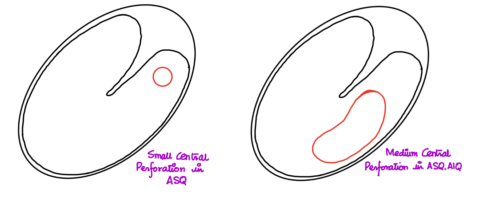
1- Small Central Perforation (Anterosuperior Quadrant)
- Draw a small circle in the anterosuperior quadrant
- Represents a minor, centrally located defect
2- Medium Central Perforation (Anterior Quadrants)
- Perforation involves anterosuperior and anteroinferior quadrants
- Draw a larger circle across both anterior quadrants
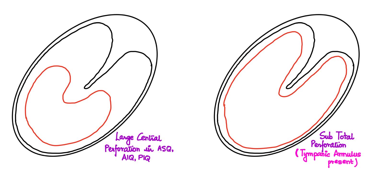
3- Large Central Perforation (Involving 3 Quadrants)
- Involves:
- Anterosuperior
- Anteroinferior
- Posteroinferior quadrants
- Not involving the annulus
4- Subtotal Perforation
- Involves almost entire pars tensa
- 🧠 Key Point: Tympanic annulus is intact
- Draw a large perforation sparing the annular rim
5- Total Perforation
- Complete loss of pars tensa including the annulus
- No tympanic membrane landmarks visible
- Draw as entire membrane missing
6- Pinhole Perforation
- Tiny perforation visible in one quadrant
- Represent with a small dot wherever located

⚠ Unsafe Types of Perforation
These are considered dangerous due to risk of cholesteatoma formation:
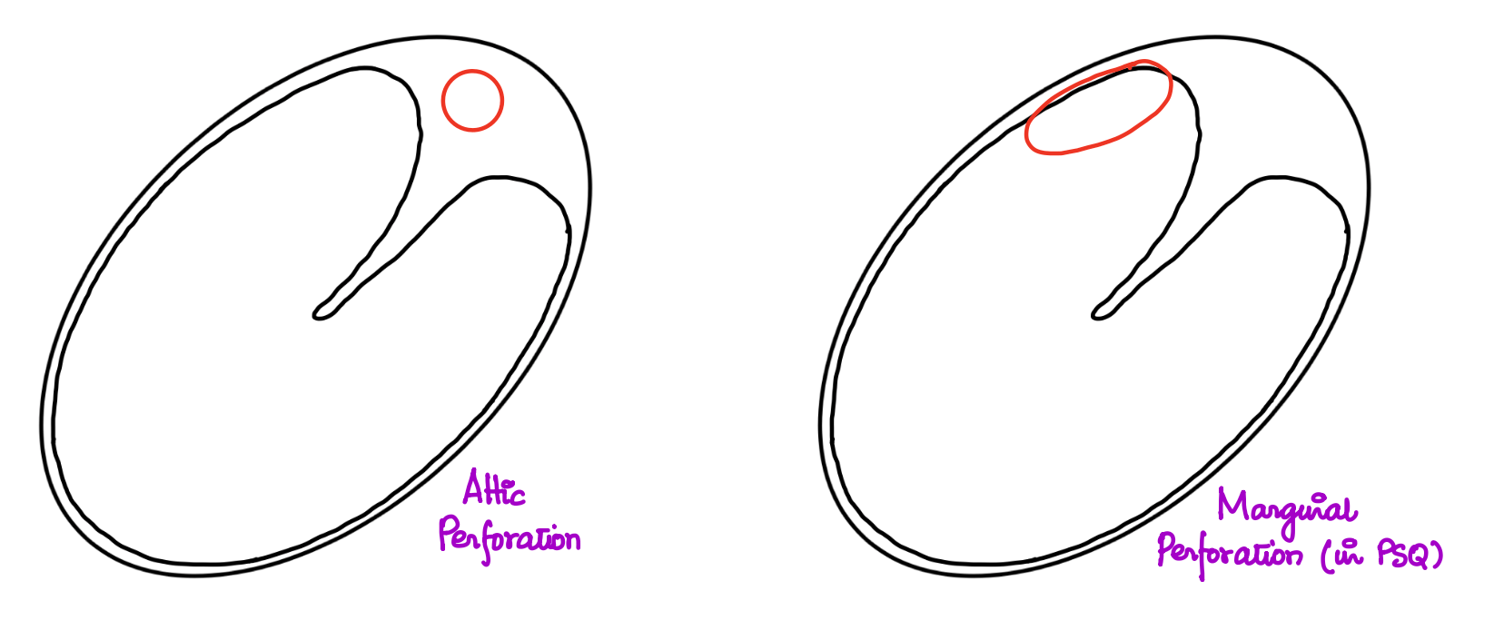
7- Attic Perforation
- Seen in the pars flaccida (above the lateral process of malleus)
- Draw in the upper region (attic area)
8- Marginal Perforation
- Edge of perforation extends up to the annulus
- Draw close to the periphery of tympanic membrane
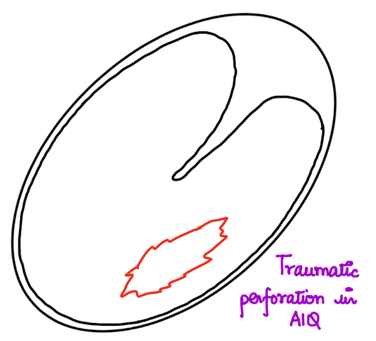
💥 Traumatic Perforation
Features:
- Ragged edges
- Often blood-stained or tinged
- Caused by:
- Barotrauma
- Slap injury
- Foreign body
- Draw irregular, jagged edges with blood tinge if needed
🔁 Multiple Perforations
- Seen in rare cases
- Draw multiple small perforations in different quadrants

🧾 Quick Summary
| Type | Key Features | Drawing Tip |
|---|---|---|
| Small Central | Limited to 1 quadrant | Small circle |
| Medium Central | Involves 2 anterior quadrants | Medium circle |
| Large Central | Involves 3 quadrants | Large circle |
| Subtotal | Nearly entire pars tensa, annulus spared | Large perforation with rim intact |
| Total | Complete membrane loss | Entire TM missing |
| Attic | Pars flaccida region | Draw at upper TM |
| Marginal | Edge touches annulus | Draw at periphery |
| Traumatic | Ragged, blood-stained | Jagged outline |
| Multiple | Multiple small defects | Multiple dots |
🎓 Final Tips
- Always mark quadrants when drawing the tympanic membrane.
- Use neat labels for clarity during the presentation.
- Know how to differentiate safe vs unsafe perforations.
📝 All topics and questions from this post are explained in detail in my Premium ENT Notes, which are designed for clinical understanding and exam success.
Residency is hard enough. Studying for it shouldn't be 😊
💎 Buy my Premium ENT Notes
Instant access to 200+ high-yield ENT notes. Your purchase includes all future updates.
🇮🇳 For Indian Students
· To buy all my notes, click here🌎 For International Students
· Buy the note for this lecture· Buy Complete Notes on Anatomy of Ear· Buy all my notes in ENT