How to Draw a Normal Tympanic Membrane
💎 Buy my Premium ENT Notes
Instant access to 200+ high-yield ENT notes. Your purchase includes all future updates.
🇮🇳 For Indian Students
· To buy all my notes, click here🌎 For International Students
· Buy the note for this lecture· Buy Complete Notes on Anatomy of Ear· Buy all my notes in ENT💡 This post is a free outline of my YouTube video. Get my full handwritten notes using the links above.
🖍 How to Draw a Normal Tympanic Membrane in Under 30 Seconds
In this post, you'll learn how to identify, draw, and label the normal tympanic membrane (TM) — an essential skill for ENT clinical exams, case presentations, and viva questions.
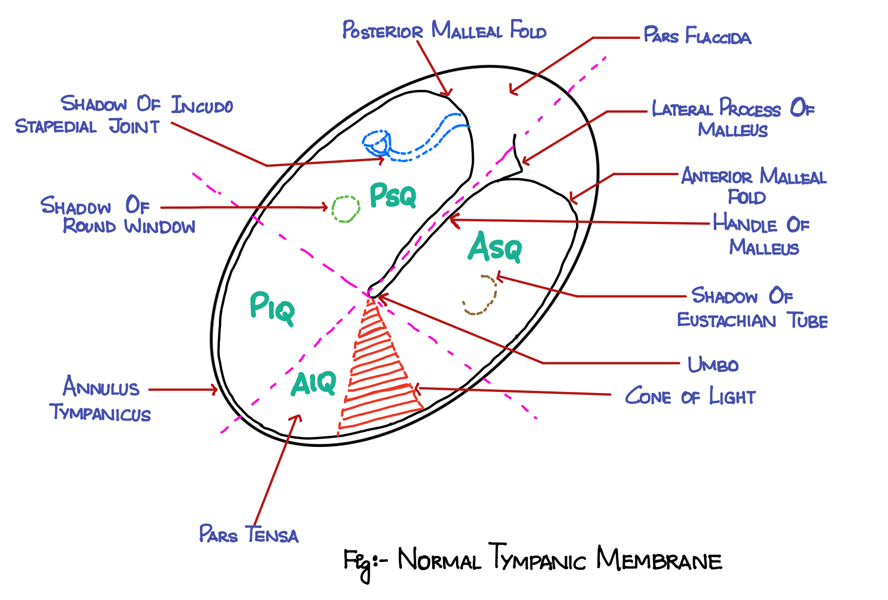
👁 What Structures Are Visible in a Normal Tympanic Membrane?
During otoscopic examination, the following structures can be visualized:
- Handle of Malleus: Long vertical shadow from upper to center
- Umbo: Tip of the handle; causes a central retraction
- Cone of Light: Seen in the anteroinferior quadrant
- Lateral Process of Malleus: Seen near the top of the handle
- Anterior & Posterior Malleal Folds: Extend from lateral process
- Pars Flaccida: Region above malleal folds
- Pars Tensa: Remaining taut portion of the membrane
- Annulus Tympanicus: Fibrocartilaginous ring surrounding the TM
🔍 Occasionally visible middle ear shadows:
- Eustachian tube shadow (anterosuperior)
- Round window (posterior superior)
- Incudostapedial joint (posterior superior)
✏️ Step-by-Step Guide to Drawing a Tympanic Membrane
Check out the YouTube video to see how to draw a normal tympanic membrane.
✅ Basic Outline:
-
Draw a tilted oval to represent the TM.
-
For the right ear, tilt right; for the left ear, tilt left.
-
Remember, the TM is angled at 45–55° to the horizontal.
✅ Add Key Landmarks:
-
Annulus Tympanicus: Outline the rim of the membrane.
-
Handle of Malleus: Long line from top center to umbo.
-
Lateral Process of Malleus: Small dot or bulge near the top.
-
Malleal Folds: Extend from lateral process to annulus (anterior & posterior).
-
Cone of Light: Wedge shape from umbo downwards.
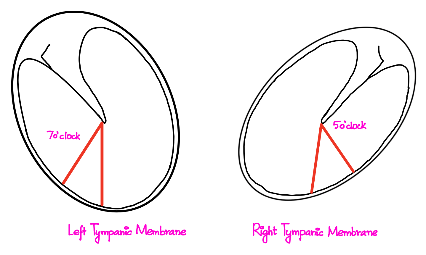
🧠 Mnemonic for Cone of Light:
- Right ear = 5 o'clock position
- Left ear = 7 o'clock position
🔲 Quadrants of Tympanic Membrane
Divide the TM into 4 quadrants for clinical documentation:
| Structure | Description |
|---|---|
| Handle of Malleus | Long vertical shadow |
| Umbo | Tip of malleus handle |
| Cone of Light | Anteroinferior quadrant |
| Lateral Process | Bulge near top of malleus |
| Malleal Folds | Form pars flaccida boundary |
| Pars Flaccida | Above malleal folds |
| Pars Tensa | Rest of the TM |
| Annulus Tympanicus | Fibrous ring around TM |
✂️ How to Divide It:
-
Draw a line along the handle of malleus (long axis).
-
Draw a perpendicular line at the umbo.
This creates 4 clear quadrants for locating perforations or pathology.
📸 Real Image Correlation
In endoscopic views, compare your drawing to real TM appearances:
-
Right TM: Tilted right, cone of light at 5 o'clock.
-
Left TM: Tilted left, cone of light at 7 o'clock.
-
Landmarks such as umbo, malleus, malleal folds, and pars tensa/flaccida remain the same.
🧾 Summary Table
| Structure | Description |
|---|---|
| Handle of Malleus | Long vertical shadow |
| Umbo | Tip of malleus handle |
| Cone of Light | Anteroinferior quadrant |
| Lateral Process | Bulge near top of malleus |
| Malleal Folds | Form pars flaccida boundary |
| Pars Flaccida | Above malleal folds |
| Pars Tensa | Rest of the TM |
| Annulus Tympanicus | Fibrous ring around TM |
🎓 Final Tips
- Practice drawing both right and left tympanic membranes.
- Always label major structures in your schematic.
- Use this skill to draw perforations accurately during long case exams.
📝 All topics and questions from this post are explained in detail in my Premium ENT Notes, which are designed for clinical understanding and exam success.
Residency is hard enough. Studying for it shouldn't be 😊
💎 Buy my Premium ENT Notes
Instant access to 200+ high-yield ENT notes. Your purchase includes all future updates.
🇮🇳 For Indian Students
· To buy all my notes, click here🌎 For International Students
· Buy the note for this lecture· Buy Complete Notes on Anatomy of Ear· Buy all my notes in ENT