Relations of Hyoglossus Muscle
💎 Buy my Premium ENT Notes
Instant access to 200+ high-yield ENT notes. Your purchase includes all future updates.
🇮🇳 For Indian Students
· To buy all my notes, click here🌎 For International Students
· Buy the note for this lecture· Buy Complete Notes on Anatomy & Embryology of Tongue· Buy all my notes in ENT💡 This post is a free outline of my YouTube video. Get my full handwritten notes using the links above.
Relations of Hyoglossus Muscle
The relations of the hyoglossus muscle is an important short question topic for your anatomy theory exams and can also come up in viva, especially in cases involving the tongue.
These relations are best understood when divided into two categories:
- Superficial (Lateral) Relations
- Deep (Medial) Relations
🔍 Deep (Medial) Relations of Hyoglossus
These are the structures deep to the hyoglossus muscle, i.e., on its medial side. Though not all are easily visualized, it's important to remember them in relation to the muscle’s origin and insertion.
🧠 Tip: You might need to memorize these as they are not as easily seen during dissection or visualized.
✅ List of Deep/Medial Relations:
- Inferior longitudinal muscle of the tongue (near the insertion)
- Middle constrictor of pharynx
- Second part of the lingual artery (close to origin)
- Stylopharyngeus muscle
- Glossopharyngeal nerve
- Stylohyoid ligament
- Junction of 1st and 2nd parts of the lingual artery (deep to posterior border of hyoglossus)
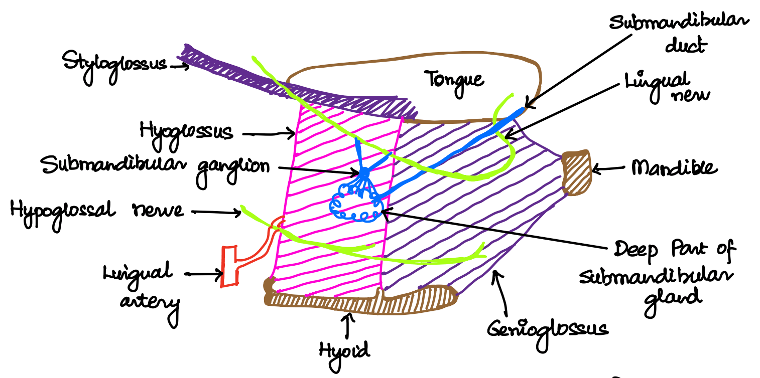
🧊 Superficial (Lateral) Relations of Hyoglossus
These are the structures lateral to the hyoglossus, lying between the hyoglossus and the mylohyoid muscle.
🎯 Remember: You can understand these better if you think of them from above downwards, as they pass in that direction between the two muscles.
📌 Important: Always draw a diagram when answering this in exams. It helps with better recall and shows the examiner your conceptual clarity.
🔽 From Above Downwards:
- Mucous membrane of the side of the tongue
- Styloglossus muscle
- Lingual nerve (usually seen in green in diagrams)
- Submandibular ganglion
- Suspended from the lingual nerve by two roots
- Deep part of the submandibular gland and submandibular duct
- Hypoglossal nerve
- Accompanied by a pair of veins
- Suprahyoid branch of the 1st part of the lingual artery
📝 All topics and questions from this post are explained in detail in my Premium ENT Notes, which are designed for clinical understanding and exam success.
Residency is hard enough. Studying for it shouldn't be 😊
💎 Buy my Premium ENT Notes
Instant access to 200+ high-yield ENT notes. Your purchase includes all future updates.
🇮🇳 For Indian Students
· To buy all my notes, click here🌎 For International Students
· Buy the note for this lecture· Buy Complete Notes on Anatomy & Embryology of Tongue· Buy all my notes in ENT