Tos Classification - Pars Flaccida Retraction
💎 Buy my Premium ENT Notes
Instant access to 200+ high-yield ENT notes. Your purchase includes all future updates.
🇮🇳 For Indian Students
· To buy all my notes, click here💡 This post is a free outline of my YouTube video. Get my full handwritten notes using the links above.
👂 Tos Classification of Pars Flaccida Retraction
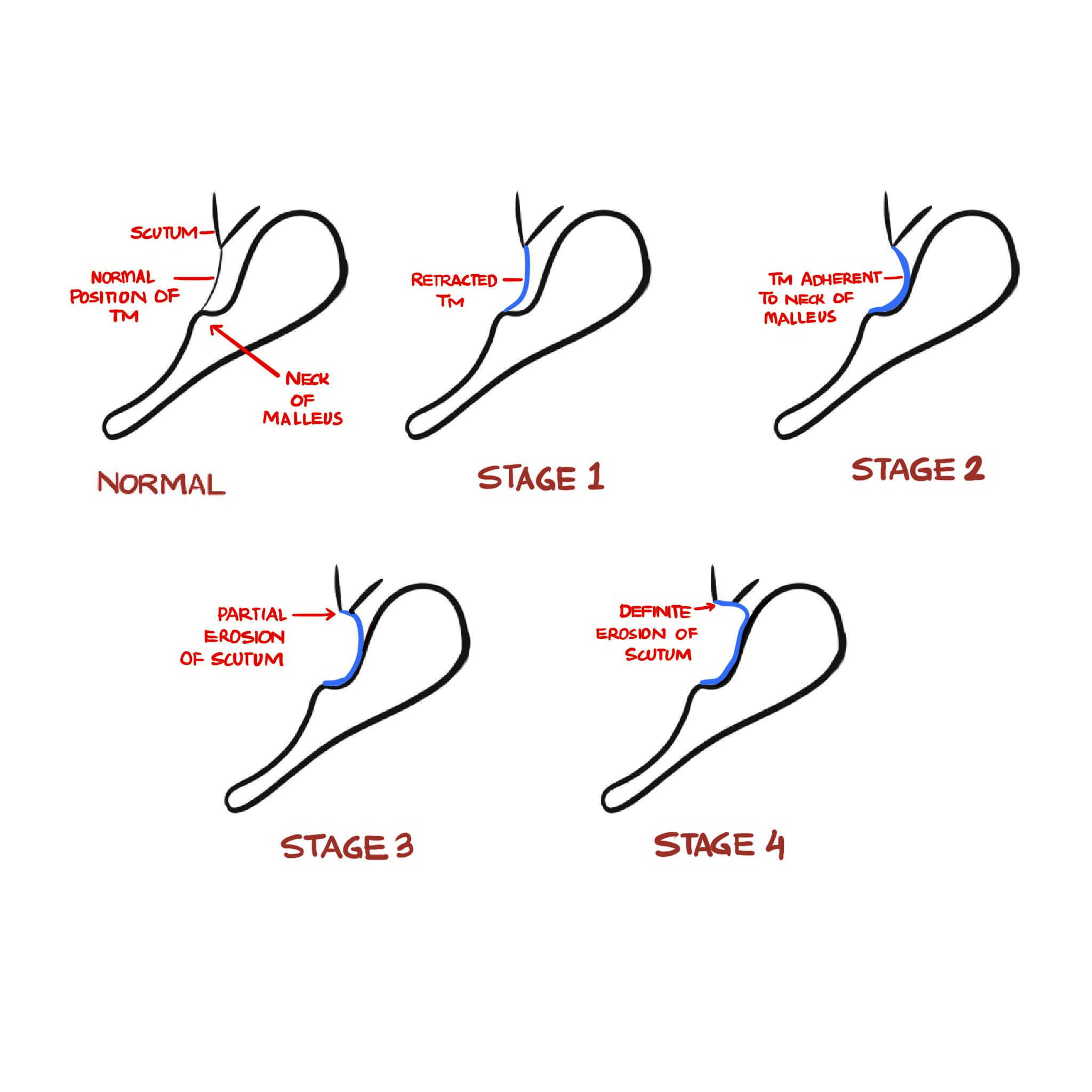
✅ Stage 1: Mild Retraction
- Minimal dimpling of pars flaccida.
- Not in contact with the neck of malleus.
- Retraction is visible but not adherent.
✅ Stage 2: Contact with Neck of Malleus
- Pars flaccida is retracted and adherent to the neck of the malleus.
- Entire retraction pocket is visible on otoendoscopy.
✅ Stage 3: Partial Erosion of Scutum
- Part of the retraction pocket is out of view.
- There is partial erosion of the bony attic wall (scutum).
- The retraction is deeper and begins to hide in the attic.
✅ Stage 4: Severe Retraction and Erosion
- Severe erosion of the scutum.
- Retraction pocket is deep, not fully visible.
- Complete adherence to surrounding structures.
📝 Summary Table
| Stage | Key Features |
|---|---|
| 1 | Mild retraction, not touching malleus |
| 2 | Adherent to neck of malleus, fully visible pocket |
| 3 | Partial scutum erosion, pocket partly hidden |
| 4 | Severe erosion, deep non-visible pocket |
~~~~~~~~
📝 All topics and questions from this post are explained in detail in my Premium ENT Notes, which are designed for clinical understanding and exam success.
Residency is hard enough. Studying for it shouldn't be 😊
💎 Buy my Premium ENT Notes
Instant access to 200+ high-yield ENT notes. Your purchase includes all future updates.
🇮🇳 For Indian Students
· To buy all my notes, click here~~~~~~~~
Related ENT Notes & Lectures
Abscesses in relation to Mastoid
Acute Mastoiditis VS Furunculosis
Acute Necrotizing Otitis Media
Acute Otitis Media - Causes, Symptoms and Treatment
Anatomy of External Ear
Anatomy of Facial Nerve
Anatomy of Facial Nerve – Branches
Anatomy of Facial Nerve – Functional Components
Anatomy of Facial Nerve – Nuclei & Course
Anatomy of Inner Ear
Anatomy of Middle Ear - Contents
Anatomy of Middle Ear - Walls & Parts
Anatomy of Tympanic Membrane
Benign Intracranial Hypertension (Otitic Hydrocephalus)
Canal wall Down VS Canal wall Up Mastoidectomy
Cholesteatoma
Chronic Suppurative Otitis Media (CSOM)
Complications of Acute Otitis Media
Complications of CSOM
Complications of Mastoidectomy
Cortical Mastoidectomy
Extracranial Complications of CSOM
False Negative Rinne Test Explained
Glomus Tumor Signs Explained | Aquino Sign, Brown Sign, Phelps Sign & Rising Sun Sign
Gradenigo Syndrome
Grommet / Tympanostomy tube / Ventilation tube
Halo Sign Explained in ENT
How to Draw a Normal Tympanic Membrane
How to Draw Tympanic Membrane Perforations
Ice Cream Cone Sign Explained in ENT
Inner Ear fluids - Perilymph and Endolymph
Inside out VS Outside in Mastoidectomy
Intracranial Complications of CSOM
Labyrinthine Fistula
Labyrinthitis
Landmarks of Facial Nerve in Mastoid and Parotid surgeries
Malignant Otitis Externa (Skull Base Osteomyelitis)
Mastoiditis
Modified Radical Mastoidectomy
Myringotomy with Grommet Insertion
Organ of Corti – Anatomy, Structure and Clinical Relevance
Otitis Media with Effusion
Otogenic Brain Abscess
Otosclerosis Part 1 - Causes, Pathogenesis, Types, Pathology
Otosclerosis Part 2 - Symptoms, Signs, Investigations, Differential diagnosis
Otosclerosis Part 3 - Treatment
Otosclerosis Signs Explained | Schwartz sign, Carhart’s notch, Halo sign, Paracusis willisii
Perichondritis - Boxer’s Ear / Cauliflower Ear
Petrositis
Radical Mastoidectomy
Referred Pain in the Ear
Sade Classification - Pars Tensa Retraction
Sigmoid Sinus Thrombosis
Theories of Cholesteatoma - Wittmack, Habermann, Ruedi, Sade
Tympanoplasty Part 1 - Definition, Types, Grafts, Indications, Contraindications
Tympanoplasty Part 2 - Approaches, Techniques, Steps & Complications
Types of Cholesteatoma - Congenital & Acquired Cholesteatoma
Types of Tympanoplasty - Wullstein Classification
Brodsky Grading of Tonsillar Enlargement
Indirect Laryngoscopy Diagram - How to Draw and Label
Relations of Hyoglossus Muscle