Sade Classification - Pars Tensa Retraction
💎 Buy my Premium ENT Notes
Instant access to 200+ high-yield ENT notes. Your purchase includes all future updates.
🇮🇳 For Indian Students
· To buy all my notes, click here💡 This post is a free outline of my YouTube video. Get my full handwritten notes using the links above.
🩺 Sade's Classification of Pars Tensa Retraction
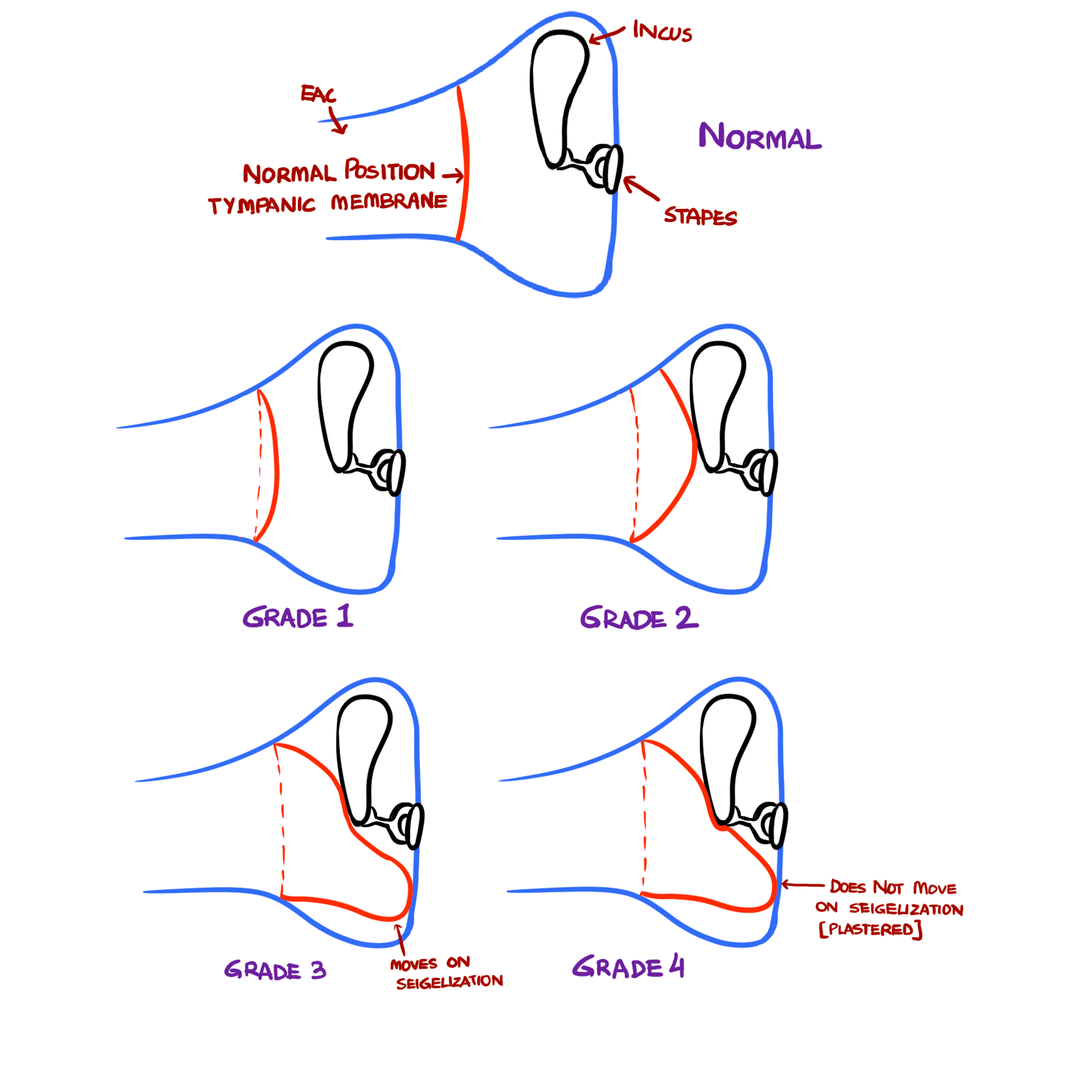
✅ Grade 1: Mild Retraction
- Minimal retraction of the tympanic membrane.
- Not in contact with the incus or any other middle ear structures.
- Loss of light reflex seen.
- Tympanic membrane still mobile.
✅ Grade 2: Contact with Ossicles
- Tympanic membrane now touches the long process of incus or stapes.
- Ossicular structures may appear more prominent on endoscopy.
✅ Grade 3: Middle Ear Atelectasis
- Retraction becomes severe, and the tympanic membrane is now in contact with the promontory.
- Key feature: The membrane still moves with Valsalva or suctioning (reversible atelectasis).
✅ Grade 4: Adhesive Otitis Media
- Tympanic membrane is plastered to the promontory.
- No movement on Valsalva or suctioning.
- Represents irreversible adhesive otitis media.
🔁 Grade 3 vs Grade 4 – Key Difference
| Feature | Grade 3 | Grade 4 |
|---|---|---|
| Contact | Tympanic membrane touches promontory | Tympanic membrane touches promontory |
| Movement on Valsalva | ✅ Present (moves) | ❌ Absent (plastered) |
| Also Known As | Middle ear atelectasis | Adhesive otitis media |
📌 Summary
The SADE classification gives insight into the severity and reversibility of pars tensa retraction:
- Early grades are mild and reversible.
- Later grades indicate more severe, potentially permanent damage.
Understanding and being able to draw and explain each stage is crucial for clinical practice and ENT exams.
~~~~~~~~
📝 All topics and questions from this post are explained in detail in my Premium ENT Notes, which are designed for clinical understanding and exam success.
Residency is hard enough. Studying for it shouldn't be 😊
💎 Buy my Premium ENT Notes
Instant access to 200+ high-yield ENT notes. Your purchase includes all future updates.
🇮🇳 For Indian Students
· To buy all my notes, click here~~~~~~~~
Related ENT Notes & Lectures
Abscesses in relation to Mastoid
Acute Mastoiditis VS Furunculosis
Acute Necrotizing Otitis Media
Acute Otitis Media - Causes, Symptoms and Treatment
Anatomy of External Ear
Anatomy of Facial Nerve
Anatomy of Facial Nerve – Branches
Anatomy of Facial Nerve – Functional Components
Anatomy of Facial Nerve – Nuclei & Course
Anatomy of Inner Ear
Anatomy of Middle Ear - Contents
Anatomy of Middle Ear - Walls & Parts
Anatomy of Tympanic Membrane
Benign Intracranial Hypertension (Otitic Hydrocephalus)
Canal wall Down VS Canal wall Up Mastoidectomy
Cholesteatoma
Chronic Suppurative Otitis Media (CSOM)
Complications of Acute Otitis Media
Complications of CSOM
Complications of Mastoidectomy
Cortical Mastoidectomy
Extracranial Complications of CSOM
False Negative Rinne Test Explained
Glomus Tumor Signs Explained | Aquino Sign, Brown Sign, Phelps Sign & Rising Sun Sign
Gradenigo Syndrome
Grommet / Tympanostomy tube / Ventilation tube
Halo Sign Explained in ENT
How to Draw a Normal Tympanic Membrane
How to Draw Tympanic Membrane Perforations
Ice Cream Cone Sign Explained in ENT
Inner Ear fluids - Perilymph and Endolymph
Inside out VS Outside in Mastoidectomy
Intracranial Complications of CSOM
Labyrinthine Fistula
Labyrinthitis
Landmarks of Facial Nerve in Mastoid and Parotid surgeries
Malignant Otitis Externa (Skull Base Osteomyelitis)
Mastoiditis
Modified Radical Mastoidectomy
Myringotomy with Grommet Insertion
Organ of Corti – Anatomy, Structure and Clinical Relevance
Otitis Media with Effusion
Otogenic Brain Abscess
Otosclerosis Part 1 - Causes, Pathogenesis, Types, Pathology
Otosclerosis Part 2 - Symptoms, Signs, Investigations, Differential diagnosis
Otosclerosis Part 3 - Treatment
Otosclerosis Signs Explained | Schwartz sign, Carhart’s notch, Halo sign, Paracusis willisii
Perichondritis - Boxer’s Ear / Cauliflower Ear
Petrositis
Radical Mastoidectomy
Referred Pain in the Ear
Sigmoid Sinus Thrombosis
Theories of Cholesteatoma - Wittmack, Habermann, Ruedi, Sade
Tos Classification - Pars Flaccida Retraction
Tympanoplasty Part 1 - Definition, Types, Grafts, Indications, Contraindications
Tympanoplasty Part 2 - Approaches, Techniques, Steps & Complications
Types of Cholesteatoma - Congenital & Acquired Cholesteatoma
Types of Tympanoplasty - Wullstein Classification
Brodsky Grading of Tonsillar Enlargement
Indirect Laryngoscopy Diagram - How to Draw and Label
Relations of Hyoglossus Muscle