Anatomy of Tympanic Membrane
💎 Buy my Premium ENT Notes
Instant access to 200+ high-yield ENT notes. Your purchase includes all future updates.
🇮🇳 For Indian Students
· To buy all my notes, click here🌎 For International Students
· Buy the note for this lecture· Buy Complete Notes on Anatomy of Ear· Buy all my notes in ENT💡 This post is a free outline of my YouTube video. Get my full handwritten notes using the links above.
Anatomy of the Tympanic Membrane
In this post, we’ll be discussing the anatomy of the tympanic membrane, which is the partition between the external ear and the middle ear.
The tympanic membrane plays a crucial role in hearing, and understanding its structure is essential for anyone studying ENT (Ear, Nose, and Throat) anatomy.
👂 Overview of the Tympanic Membrane
The tympanic membrane is located at the medial end of the external auditory canal (EAC), forming the majority of the lateral wall of the tympanic cavity.
It is oval in shape, with a broader superior aspect than inferior.
Dimensions:
- Height: 9-10 mm
- Width: 8-9 mm
- Thickness: 0.1 mm
The tympanic membrane is positioned obliquely, forming an angle of approximately 55° with the deep EAC.
This angle and the slightly lateral position of the posterior superior part of the tympanic membrane give it a characteristic appearance.
🔬 Parts of the Tympanic Membrane
The tympanic membrane consists of two primary parts:
- Pars Tensa
- Pars Flaccida
1- Pars Tensa
The pars tensa makes up most of the tympanic membrane and is the stiff, tense portion. It is visible as the grey shaded area in images of the tympanic membrane.
- Annulus Tympanicus
At the periphery of the pars tensa, the tympanic membrane becomes thickened, forming a fibrocartilaginous ring called the annulus tympanicus. This ring fits into a groove in the bony meatal canal, known as the tympanic sulcus.
-
Umbo is the central portion of the tympanic membrane near the tip of the malleus, where the membrane is slightly tented inward.
-
The cone of light, visible during a clinical exam, radiates from the umbo, indicating a normal tympanic membrane. This feature may be lost in cases of pathology.
2- Pars Flaccida
The pars flaccida is located above the lateral process of the malleus. This region is less tense than the pars tensa and often appears slightly pinkish.
What is Pars Flaccida also known as?
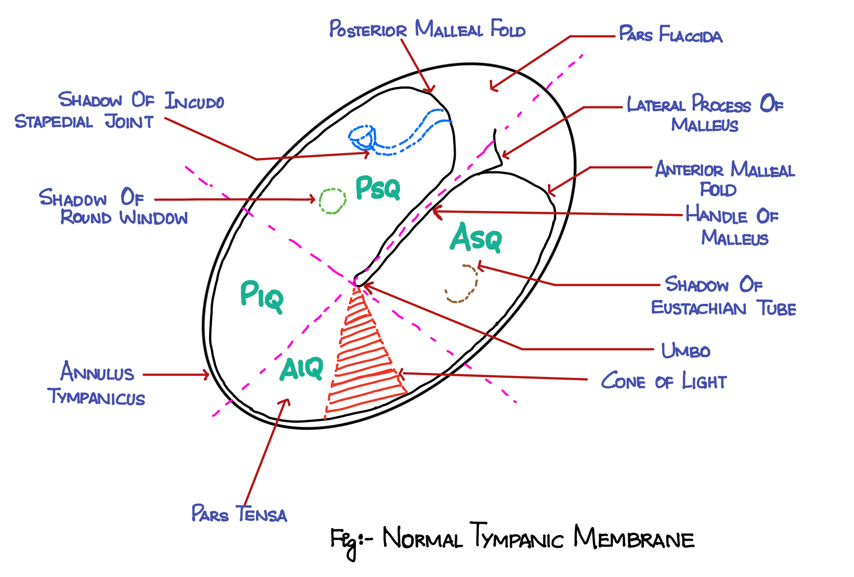
📌 Key Landmarks of the Tympanic Membrane
While examining the tympanic membrane, certain landmarks are important to note:
- Lateral Process of the Malleus
- Handle of the Malleus
- Umbo
- Cone of Light (anterior inferior quadrant)
- Anterior and Posterior Malleal Folds
- Incus Shadow (may or may not be visible)
- Tympanic Annulus
🧬 Layers of the Tympanic Membrane
The tympanic membrane has three distinct layers:
-
Outer Epithelial Layer: Continuous with the skin of the external auditory canal (EAC).
-
Middle Fibrous Layer: Contains three types of fibers:
- Radial fibers
- Circular fibers
- Parabolic fibers
-
Innermost Mucosal Layer: Continuous with the middle ear mucosa.
🩸 Blood Supply of Tympanic Membrane
The blood supply to the tympanic membrane comes from the same sources that supply both the external auditory meatus and the middle ear.
These vessels form an anastomosis within the connective tissue layer of the lamina propria (the middle fibrous layer).
The main blood vessels involved include:
-
Epidermal Vessels: Arising from the deep auricular branch of the maxillary artery.
-
Mucosal Vessels: Arising from the anterior tympanic branch of the maxillary artery, stylomastoid branch of the posterior auricular artery, and possibly the middle meningeal artery.
⚡ Nerve Supply of Tympanic Membrane
The nerve supply of the tympanic membrane is provided by three main nerves:
-
Auriculotemporal Nerve: A branch of the mandibular division of the trigeminal nerve. It supplies the anterior half of the lateral surface of the tympanic membrane.
-
Auricular Branch of the Vagus Nerve: Supplies the posterior half of the lateral surface of the tympanic membrane.
What is the other name for Auricular branch of Vagus nerve?
- Tympanic Branch of the Glossopharyngeal Nerve: Supplies the medial surface of the tympanic membrane.
What is the other name for Tympanic Branch of the Glossopharyngeal Nerve?
👨⚕️ Clinical Examination of the Tympanic Membrane
During a clinical examination, certain features are essential to observe:
- Cone of Light (Anterior Inferior Quadrant)
- Umbo (At the center near the malleus)
- Handle of the Malleus (Visible as a bony structure)
- Lateral Process of the Malleus
- Anterior and Posterior Malleal Folds
- Incus Shadow (Can be visible, depending on the patient's anatomy)
- Tympanic Annulus (The peripheral thickened ring)
🎓 Conclusion
Understanding the anatomy of the tympanic membrane is crucial for clinical ENT practice, as it helps in diagnosing various ear conditions. Familiarizing yourself with the structure, landmarks, layers, blood supply, and nerve supply of the tympanic membrane will enable you to make informed clinical observations and interpretations.
If you'd like to learn how to draw the tympanic membrane easily, check out my post on drawing the tympanic membrane in 30 seconds.
📝 All topics and questions from this post are explained in detail in my Premium ENT Notes, which are designed for clinical understanding and exam success.
Residency is hard enough. Studying for it shouldn't be 😊
💎 Buy my Premium ENT Notes
Instant access to 200+ high-yield ENT notes. Your purchase includes all future updates.
🇮🇳 For Indian Students
· To buy all my notes, click here🌎 For International Students
· Buy the note for this lecture· Buy Complete Notes on Anatomy of Ear· Buy all my notes in ENT